Support and Learning Resources for Young Arthritis Patients
Close to 300,000 young individuals in the U.S. are affected by juvenile idiopathic arthritis (JIA) and related pediatric rheumatic conditions. These autoimmune disorders can impact joints, skin, eyes, and even internal organs. While receiving such a diagnosis might feel overwhelming, it's comforting to know that there are effective treatments to help manage the condition.
Juvenile arthritis encompasses a range of rheumatic conditions that affect children 16 years and younger. It's important to note that these aren't simply adult diseases appearing in kids; they have unique characteristics and require different treatment approaches. Among these conditions, juvenile idiopathic arthritis (formerly known as juvenile rheumatoid arthritis) is the most prevalent. Other examples include juvenile psoriatic arthritis, pediatric lupus, and several more.

How New Imaging Technology is Changing Arthritis Diagnosis and Treatment
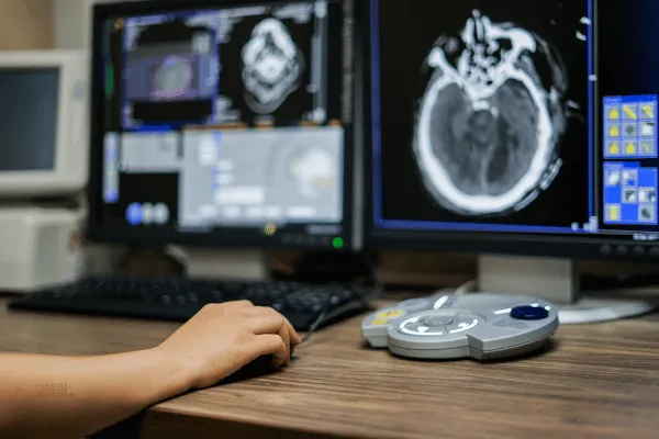
What if doctors could diagnose arthritis earlier, tell the difference between types of arthritis more accurately, and figure out how well treatments are working—all with better imaging? That’s exactly what’s happening with today’s advancements in medical imaging.
At a recent arthritis conference, Dr. Georg Schett, a leading expert from Friedrich-Alexander University in Germany, explained how new imaging tools are helping doctors better understand inflammatory arthritis. Let’s break down the highlights and what they mean for people living with arthritis.
Why Imaging Matters for Arthritis
Arthritis isn’t just one disease—it includes many conditions, like rheumatoid arthritis (RA) and psoriatic arthritis (PsA). Each condition affects the body differently, so diagnosing them accurately is very important.
Dr. Schett shared how new imaging methods, like MRIs and CT scans, combined with artificial intelligence (AI), can help doctors spot specific patterns of inflammation unique to each type of arthritis. These tools even help catch arthritis early, giving patients a head start on treatment.
“The more data we give these systems, the better they can identify the differences,” Dr. Schett explained.
Why Traditional X-Rays Aren’t Enough
For years, doctors have used X-rays to look at arthritis, but X-rays don’t always show the full picture—especially in the early stages of the disease.
That’s where newer tools like 3D imaging and high-resolution CT scans come in. These techniques provide a clearer view of bone and joint changes, allowing doctors to see the damage more accurately.
“CT scans can also check for joint damage, like erosions, and monitor how well treatments are working,” Dr. Schett said.
How Imaging Can Look Inside Joints
One of the most exciting advancements is something called functional imaging. While traditional scans show what the joint looks like, functional imaging can show how the joint is working. For example, it can measure oxygen levels or how much water is in the tissue.
A new technology called multispectral optoacoustic tomography (MSOT) combines lasers and ultrasound to detect changes in the joint’s metabolism (how the joint tissues use energy). This could help doctors predict which psoriasis patients are more likely to develop psoriatic arthritis.
Learning How Inflammation Affects the Body
Another focus of new imaging is understanding how inflammation spreads in the body. For example, a type of cell called fibroblasts plays a role in inflammation. These cells are found in both RA and certain cancers.
Doctors can now use advanced scans, like PET scans, to track these fibroblasts and see how well treatments are working. While this technique is mostly used in cancer treatment, Dr. Schett believes it could also improve arthritis care.
“We can learn a lot about how inflammation progresses and which therapies might work best,” he explained.
What Does This Mean for People with Arthritis?
These advancements in imaging technology mean better care for people with arthritis. Here’s why:
Earlier Diagnosis: Doctors can catch arthritis earlier, which gives patients a better chance to manage the disease and slow its progression.
More Accurate Diagnoses: New imaging tools can tell the difference between types of arthritis more clearly, so treatments can be more targeted.
Improved Treatment Monitoring: By seeing how joints and tissues respond to treatment, doctors can adjust therapies to make them more effective.
Looking Ahead
These new imaging tools are opening up exciting possibilities in arthritis care. At the American Arthritis Foundation, we’re committed to sharing these advancements and supporting research that improves lives.
If you’ve experienced advanced imaging as part of your arthritis care, we’d love to hear your story! Let’s work together to spread awareness about these breakthroughs and keep pushing for better treatments for everyone with arthritis.
Follow us for more updates on how technology is transforming arthritis care.
Have a question?
We're Here to Help
By providing my phone number, I agree to receive text messages from the business.

